42 neuron structure labeled
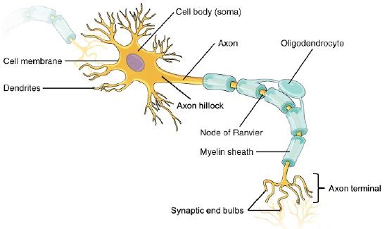
Neuron under Microscope with Labeled Diagram - AnatomyLearner The neuron structure has two main components: the cell body and the neuron processes (axons and dendrite). Let's see the neuron histology slide labelled diagram and try to find out the below-mentioned characteristics - Presence of an identifiable cell body (soma) that locates in the brain's grey matter (according to the slide image). Neuron: Definition, Structure, Diagram, Parts & Functions - Collegedunia The neuron is a highly specialized cell that is responsible for the transmission of nerve impulses. It is classified into unipolar, bipolar and multipolar neurons. On the basis of transmission mechanism, neurons are classified into sensory, motor and interneurons. Dendrites, Cell body and Axon are the main parts of the neuron.
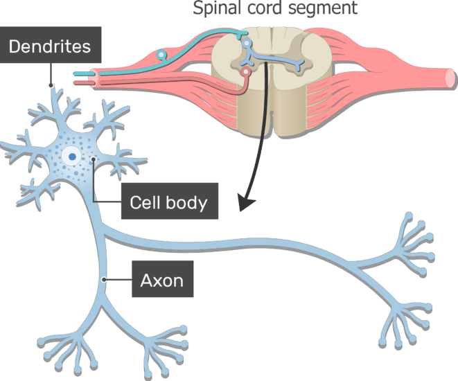
Overview of neuron structure and function - Khan Academy These include nerve cells (or neurons) and glial cells (or glia ). Neurons are the basic functional units of the nervous system, and they generate electrical signals called action potentials, which allow them to quickly transmit information over long distances.

Neuron structure labeled
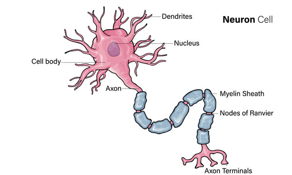
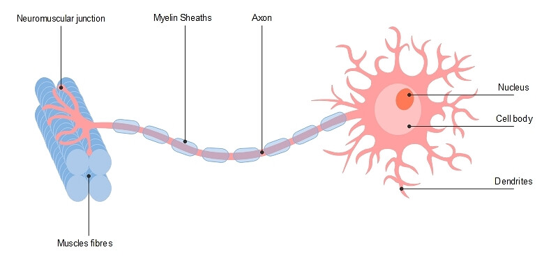
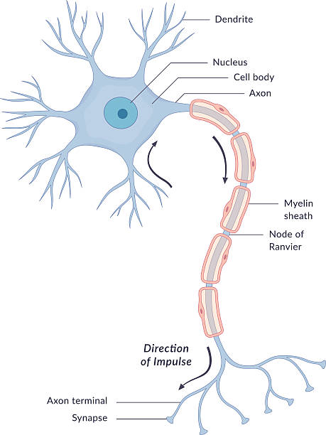
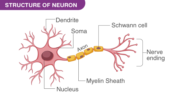
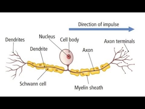
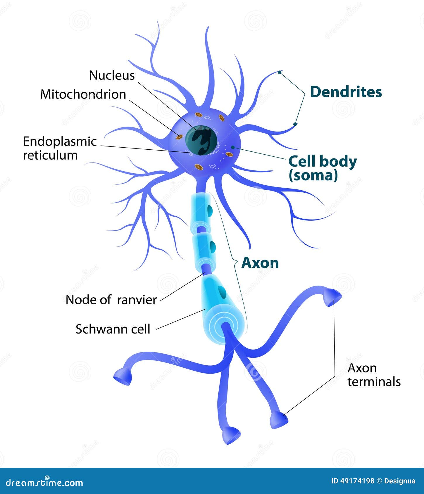
Parts of a Neuron and Their Functions with Labelled Diagram - Science Facts While they have the common features of a typical cell, they are structurally and functionally unique from other cells in many ways. All neurons have three main parts: 1) dendrites , 2) cell body or soma, and 3) axons. Besides the three major parts, there is the presence of axon terminal and synapse at the end of the neuron. en.wikipedia.org › wiki › Pyramidal_cellPyramidal cell - Wikipedia The large amount of branching allows the neuron to send and receive signals to and from many different neurons. Pyramidal neurons, like other neurons, have numerous voltage-gated ion channels . In pyramidal cells, there is an abundance of Na + , Ca 2+ , and K + channels in the dendrites, and some channels in the soma. A Guide to Understand Neuron with Neuron Diagram The main structure of a neuron includes the following parts: Dendrite: The cell body may have some branch-like structures, which work in receiving the signals. They are known as dendrite. A neuron may have multiple dendrites, while some of them may not have any. The dendrites receive signals from other neurons and pass them on to the cell body.
Neuron structure labeled. Animal Cell- Definition, Structure, Parts, Functions, Labeled Diagram Web17.09.2022 · Animal cell size and shape. Animal cells come in all kinds of shapes and sizes, with their size ranging from a few millimeters to micrometers. The largest animal cell is the ostrich egg which has a 5-inch diameter, weighing about 1.2-1.4 kg and the smallest animal cells are neurons of about 100 microns in diameter. Neuron Diagram & Types | Ask A Biologist - Arizona State University Unipolar neurons are also known as sensory neurons. They have one axon and one dendrite branching off in opposite directions from the cell body. These cells pass signals from the outside of your body, such as touch, along to the central nervous system. Bipolar neurons have one axon and only one dendrite branch. en.wikipedia.org › wiki › Purkinje_cellPurkinje cell - Wikipedia Parallel fibers pass orthogonally through the Purkinje neuron's dendritic arbor, with up to 200,000 parallel fibers forming a Granule-cell-Purkinje-cell synapse with a single Purkinje cell. Each Purkinje cell receives approximately 500 climbing fiber synapses, all originating from a single climbing fiber. A Labelled Diagram Of Neuron with Detailed Explanations - BYJUS A neuron is also known as the nerve cell. The structure of a neuron varies with their shape and size and it mainly depends upon their functions and their location. Neurons are the structural and functional units of the nervous system. A group of neurons forms a nerve. Neurons are the structural and functional units of the nervous system.
› searchSearch NCBI databases - NLM Search all biomedical databases provided by the National Center for Biotechnology Information (NCBI), an agency of the U.S. National Library of Medicine at the NIH Diagram Quiz on Neuron Structure and Function (Labeling Quiz) 1. Identify the cell type in the above figure Liver Cell Cardiac Cell Nerve cell Skin cell 2. In the figure, labeled '1' receives impulses from adjacent neuron. It is called the Dendron Dendrite Axon Axonite 3. In the figure, labeled '2' is the short filaments from the cell body that carries impulses from dendrites to the cell body which is the A Labelled Diagram of Neuron with Detailed decription - Collegedunia A neuron is a type of cell that is largely responsible for conveying information via electrical and chemical impulses. The brain, spinal cord, and peripheral nerves all contain them. The nerve cell is another name for a neuron. The structure of a neuron changes depending on its form and size, as well as its function and location. Neuron Parts, Structure, & Function | What is a Neuron? - Video ... Neuron structure is very important for its function because neurons are specifically designed to transmit electrical signals from cell to cell. The soma is the body of the neuron and is also...
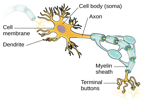
Label Parts of a Neuron Diagram | Quizlet Dendrites. receives impulses from other nerve cells. axon hillock. The cell body...the part of the cell that houses the nucleus and keeps the entire cell alive and functioning. Myelin Sheath. Surrounds the axon an insulates it from surrounding cells and tissues and making signal transitions faster and more efficient. Terminal Buttons. What Is a Neuron? - Definition, Structure, Parts and Function - BYJUS Neurons are the building blocks of the nervous system. They receive and transmit signals to different parts of the body. This is carried out in both physical and electrical forms. There are several different types of neurons that facilitate the transmission of information. EE Times - Connecting The Global Electronics Community WebEE Times offers reliable electronics news, engineering resources, podcasts, papers, and events from Award-winning journalists. Visit to learn more. Neurons (With Diagram) - Biology Discussion A neuron is a structural and functional unit of the neural tissue and hence the neural system. Certain neurons may almost equal the length of body itself. Thus neurons with longer processes (projections) are the longest cells in the body. Human neural system has about 100 billion neurons. Majority of the neurons occur in the brain.
Neurons | Organismal Biology
Labeled Neuron Diagram - Science Trends Neurons can be differentiated based on where they originate, where they connect to, their anatomical structure, and what function they perform in the nervous system. In general, there are a handful or structural varieties of neurons: The structural varieties of neurons. Credit: CNX OpenStax Biology via WikiCommons CC-BY 4.0
11.3: Neurons - Biology LibreTexts
Nervous System - Label the Neuron - TheInspiredInstructor.com Nervous System - Neuron: Nerve Cell. Choose the correct names for the parts of the neuron. (6) This neuron part receives messages from other neurons. (7) This neuron part sends on messages to other neurons. (8) This neuron part gives messages to muscle tissue. (9) This neuron part processes incoming messages.
Neuron Structure" Images – Browse 13 Stock Photos, Vectors ...
› products › by-typeInfinium Global Screening Array-24 Kit | Population-scale ... Global Content. The Infinium Global Screening Array-24 BeadChip combines multi-ethnic genome-wide content, curated clinical research variants, and quality control (QC) markers for precision medicine research.
A Guide to Understand Neuron with Neuron Diagram | EdrawMax ...
What Is a Neuron? Diagrams, Types, Function, and More - Healthline Neurons, also known as nerve cells, send and receive signals from your brain. While neurons have a lot in common with other types of cells, they're structurally and functionally unique....
Neuron Diagram Stock Illustration - Download Image Now ...
The Neuron - External Structure and Classification | Interactive ... There are 3 different types of neurons based on their structure: Multipolar neurons: one axon, many dendrites Bipolar neurons: one axon, one dendrite Unipolar neurons: single axon Pseudounipolar neurons: One axon that branches in two. That's it for this introduction to the neuron's structure.

Draw neat labeled diagrams of neurons
Search NCBI databases - NLM WebStructure. Experimentally-determined biomolecular structures. BLAST. A tool to find regions of similarity between biological sequences. blastn. Search nucleotide sequence databases. blastp. Search protein sequence databases. blastx. Search protein databases using a translated nucleotide query. tblastn . Search translated nucleotide databases using a …
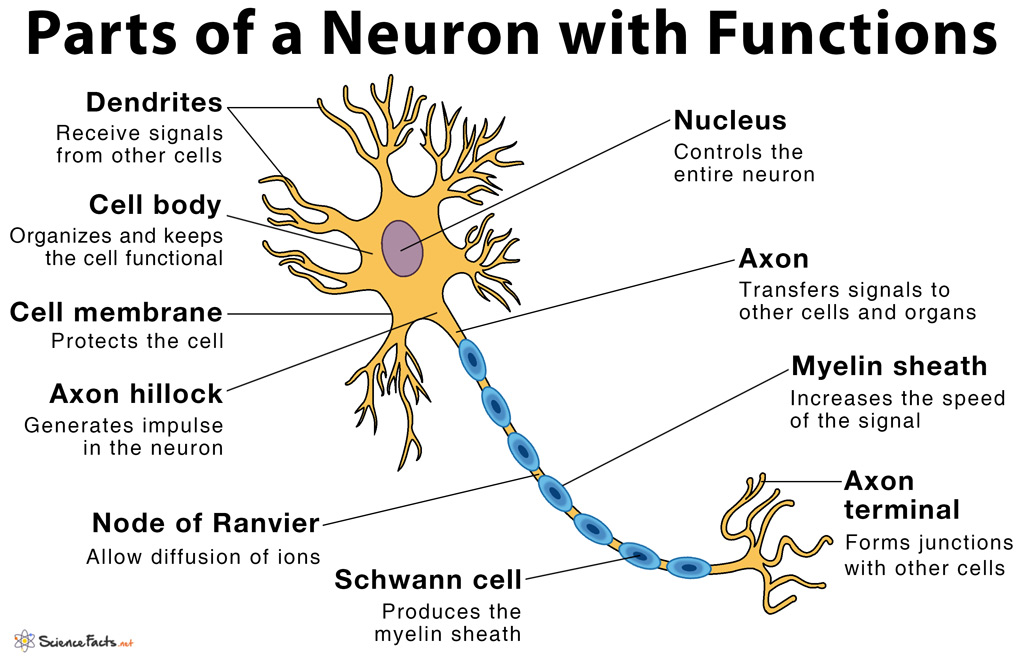
Parts of a Neuron and Their Functions with Labelled Diagram
Neurons - Structure and Classification , Anatomy QA Neurons are classified into various types : 1. According to the shape and number of processes: Pseudounipolar neuron: single process arises from the cell body which divides to form dendrite and axon ( central and peripheral process). e.g. dorsal root ganglion cells of the spinal cord. Bipolar neuron: two processes arise from the cell body, one ...

Dendrites: definition, structure and function | GetBodySmart
Neurons: Structure, Types, How They Work, Functions - Verywell Health Neurons: Structure, Types, How They Work, Functions Neurons (nerve cells) are the basic units of the nervous system. Learn more about the functions, structure, and types of neurons. Neurons (nerve cells) are the basic units of the nervous system. Learn more about the functions, structure, and types of neurons. Menu Verywell Health What Are Neurons?

Learn About Labeled Lines Of Neuron | Chegg.com
Structure of Neurons: What Is a Neuron? Types, Structure, Parts Structure of Neuron Each neuron has a cell body, which is the central area of the neuron. It contains the nucleus and other structures common to all cells in the body, such as mitochondria. Neurons have highly branched fibres that reach out from the neuron are called dendritic trees. Each branch is called a dendrite.

Neurons (Nerve Cells) Structure, Function & Types - Simply ...
Neuro-linguistic programming - Wikipedia WebNeuro-linguistic programming (NLP) is a pseudoscientific approach to communication, personal development and psychotherapy, that first appeared in Richard Bandler and John Grinder's 1975 book The Structure of Magic I.NLP claims that there is a connection between neurological processes (neuro-), language (linguistic) and acquired behavioral …

Axon - Structure and Functions | GetBodySmart
Amyloid beta: structure, biology and structure-based … Web17.07.2017 · Amyloid beta peptide (Aβ) is produced through the proteolytic processing of a transmembrane protein, amyloid precursor protein (APP), by β- and γ-secretases. Aβ accumulation in the brain is ...
Parts of a neuron labeled and neuron structure | GetBodySmart
Dopamine - Wikipedia WebStructure. A dopamine molecule consists of a catechol structure (a benzene ring with two hydroxyl side groups) with one amine group attached via an ethyl chain. As such, dopamine is the simplest possible catecholamine, a family that also includes the neurotransmitters norepinephrine and epinephrine. The presence of a benzene ring with this amine …

Human Neuron Structure Nerve Cell Medical Chart Stock Vector ...
Types of Neurons: Parts, Structure, and Function - Verywell Health Types of neurons based on structure include: Unipolar neurons: These neurons have a single long axon that is responsible for sending electrical signals. The axon in unipolar neurons is myelinated, which allows for rapid signal transmission. Multipolar neurons: These neurons are able to receive impulses from multiple neurons via dendrites.

a Draw the structure of a neuron and label the following on ...
Structure of a Neuron - Owlcation The neuron is broken up into two major regions: A region for receiving and processing incoming information from other cells A region for conducting and transmitting information to other cells The type of information that is received, processed, and transmitted by a neuron depends on its location in the nervous system.
What Is a Neuron? - Definition, Structure, Parts and Function
ufldl.stanford.edu › tutorial › unsupervisedUnsupervised Feature Learning and Deep Learning Tutorial Informally, we will think of a neuron as being “active” (or as “firing”) if its output value is close to 1, or as being “inactive” if its output value is close to 0. We would like to constrain the neurons to be inactive most of the time. This discussion assumes a sigmoid activation function.
How to draw sensory neuron well labelled and very easy
Video: Neuron Structure | Anatomy and Physiology I Video: Neuron Structure. Neuron Structure - Neuroanatomy Basics - Anatomy Tutorial.
Neuron Structure Stock Illustrations – 15,057 Neuron ...
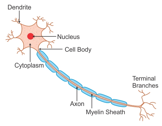
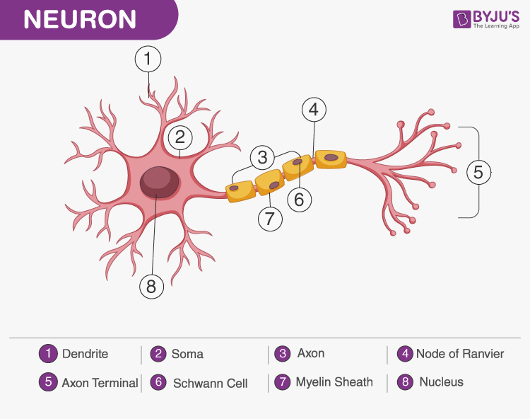
Neuron Diagram Labeled | EdrawMax Template It is an effective form of self-assessment, enabling students to check their understanding. In the following diagram, we have illustrated the important parts of the Neuron. In the following Neuron labeled diagram, we have dendrite, cell body, axon, myelin sheath, Schwann cell, a node of Ranvier, axon terminal, and nucleus.
CBSE Class 9 Answered
Neuron Structure and Classification - BIO 264 Anatomy & Physiology I Neuron Structure and Classification 6.1.2. Glial Cells of the CNS 6.1.3. Glial Cells of the PNS 6.2. PHYSIOLOGY OF THE NEURON 6.2.1. The Synapse 6.2.2. Summation 7.0. MODULE 7: SKELETAL MUSCLE 7.1. FUNCTIONS AND PROPERTIES OF SKELETAL MUSCLE TISSUE 7.2. SKELETAL MUSCLE ORGANIZATION 7.2.1. Gross and Microscopic Structure 7.3.

2: Schematic representation of the structure of a typical ...
Label a Neuron Quiz - PurposeGames.com Label a Neuron — Quiz Information. This is an online quiz called Label a Neuron. There is a printable worksheet available for download here so you can take the quiz with pen and paper. From the quiz author. Title says it all. Quiz Points. 10 p. You need to get 100% to score the 10 points available.

Structure of the Neurons The Best Definition By LTA
China Brain Project: Basic Neuroscience, Brain Diseases, and Brain ... Web02.11.2016 · The China Brain Project, entitled “Brain Science and Brain-Inspired Intelligence,” is formulated as a 15-year plan (2016–2030), with the first five years coincident with China’s 13 th five-year plan for national social and economic development. As a relatively new research discipline in China, neuroscience has a small community and …

Q1 With the help of a suitable diagram describe the ...
Neuron - Wikipedia Neurons are the primary components of the nervous system, along with the glial cells that give them structural and metabolic support. [1] The nervous system is made up of the central nervous system, which includes the brain and spinal cord, and the peripheral nervous system, which includes the autonomic and somatic nervous systems. [2]

587 Neuron Labeled Images, Stock Photos & Vectors | Shutterstock
› createJoin LiveJournal Password requirements: 6 to 30 characters long; ASCII characters only (characters found on a standard US keyboard); must contain at least 4 different symbols;

Labeled Neuron Diagram - Science Trends
Labeled Neuron Diagram Pictures, Images and Stock Photos Browse 20 labeled neuron diagram stock photos and images available, or start a new search to explore more stock photos and images. Carpal tunnel vector illustration scheme. Medical labeled... Carpal tunnel vector illustration scheme. Medical labeled diagram closeup with isolated muscle, transverse carpal ligament, median nerve, tendon sheath ...
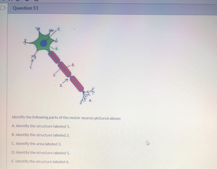
Solved D Question 51 3. Identify the following parts of the ...
› synapseSynapse | Its Structure, Types, Function and Transmission Steps Mar 29, 2021 · This is achieved through transmission through synapses. A neuron is the structural and functional unit of the nervous system. Structure of neuron. A neuron has three parts- a cell body, dendrites, and an axon ending at an axon terminal. A Neuron structure with Synapses at the end. The cell body (soma) contains the nucleus and cytoplasm.
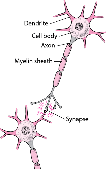
Figure: Typical Structure of a Nerve Cell - MSD Manual ...
20 Neuron Diagram Labeled Illustrations & Clip Art - iStock Browse 20 neuron diagram labeled stock illustrations and vector graphics available royalty-free, or start a new search to explore more great stock images and vector art. Newest results. The nervous system. The human nervous system vector medical illustration. Carpal tunnel vector illustration scheme.

Label the parts of a typical neuron (1, 2, 3, 4, 5, 6) shown ...
Machine Learning Glossary | Google Developers Web07.11.2022 · In that case, the neuron calculates the sigmoid of -2.0, which is approximately 0.12. Therefore, the neuron passes 0.12 (rather than -2.0) to the next layer in the neural network. The following figure illustrates the relevant part of the process: active learning. A training approach in which the algorithm chooses some of the data it learns from. Active …

Structure of a multipolar neuron | Neurons, Human anatomy and ...
Hypothalamus - Wikipedia WebThe hypothalamus (from Ancient Greek ὑπό (hupó) 'under', and θάλαμος (thálamos) 'chamber') is a part of the brain that contains a number of small nuclei with a variety of functions. One of the most important functions is to link the nervous system to the endocrine system via the pituitary gland.The hypothalamus is located below the thalamus and is …

Basic structure of a neuron. Inset shows a synaptic ...
Join LiveJournal WebPassword requirements: 6 to 30 characters long; ASCII characters only (characters found on a standard US keyboard); must contain at least 4 different symbols;

Neuron Diagram, Structure & Function | What Is a Neuron ...
An Easy Guide to Neuron Anatomy with Diagrams - SimplyPsychology.org Neurons, also known as nerve cells, are essentially the cells that make up the brain and the nervous system. Neurons do not touch each other, but where one neuron comes close to another neuron, a synapse is formed between the two. According to new research, the human brain contains around 86 billons neurons (Herculano-Houzel, 2009).
Neurons | Introduction to Psychology
A Guide to Understand Neuron with Neuron Diagram The main structure of a neuron includes the following parts: Dendrite: The cell body may have some branch-like structures, which work in receiving the signals. They are known as dendrite. A neuron may have multiple dendrites, while some of them may not have any. The dendrites receive signals from other neurons and pass them on to the cell body.

Draw a labelled diagram of a neuron with a myelin sheath.
en.wikipedia.org › wiki › Pyramidal_cellPyramidal cell - Wikipedia The large amount of branching allows the neuron to send and receive signals to and from many different neurons. Pyramidal neurons, like other neurons, have numerous voltage-gated ion channels . In pyramidal cells, there is an abundance of Na + , Ca 2+ , and K + channels in the dendrites, and some channels in the soma.
Solved 1. Identify the three major parts of a typical | Chegg.com
Parts of a Neuron and Their Functions with Labelled Diagram - Science Facts While they have the common features of a typical cell, they are structurally and functionally unique from other cells in many ways. All neurons have three main parts: 1) dendrites , 2) cell body or soma, and 3) axons. Besides the three major parts, there is the presence of axon terminal and synapse at the end of the neuron.
A Labelled Diagram Of Neuron with Detailed Explanations

Draw a labelled diagram of a neuron

Parts of a neuron labeled and neuron structure | GetBodySmart

Motor neuron | Alila Medical Images

Axon vector illustration. labeled diagram with explanation ...

Draw a labelled diagram of a neuron.

Draw the diagram of neuron and label any two parts.
Neural control and coordination | Class 11 Biology (India ...

Describe the structure of neuron with the help of a neat ...

Long answer question Draw the neat labelled diagram of ...


Komentar
Posting Komentar