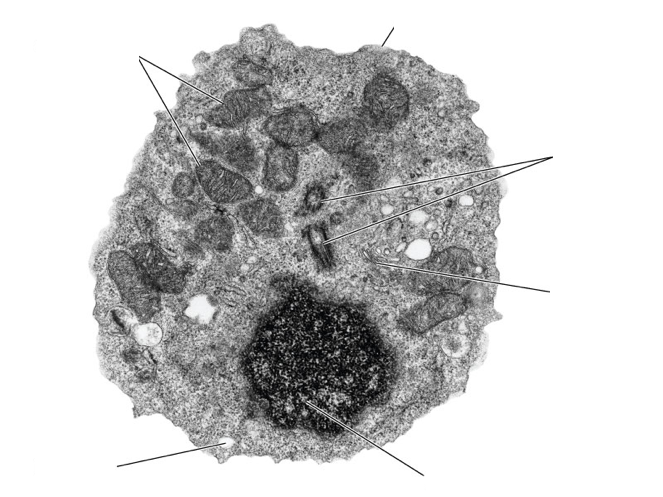
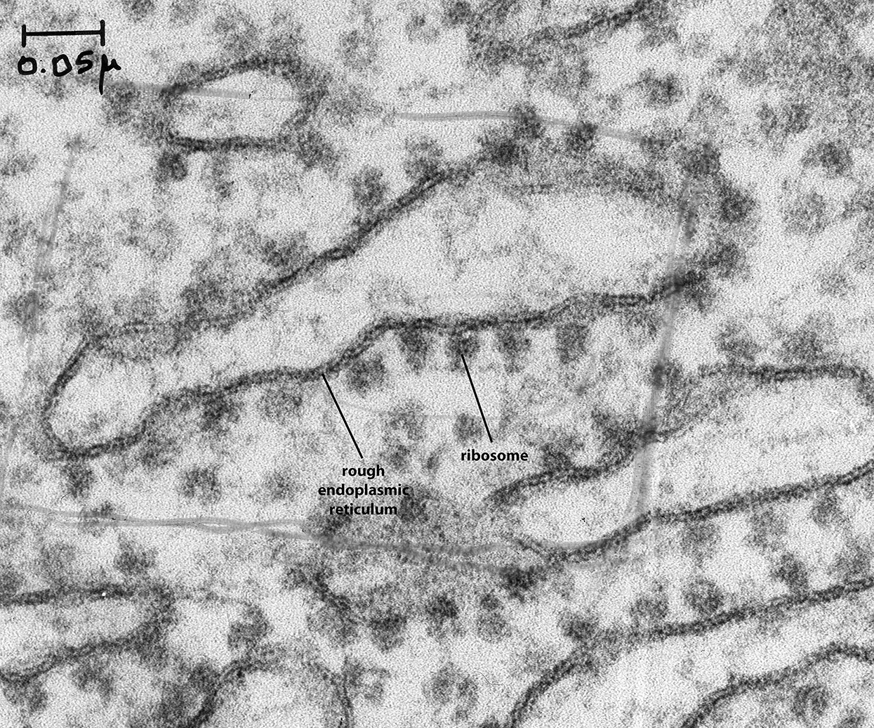
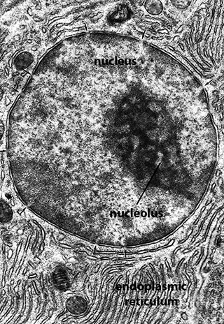
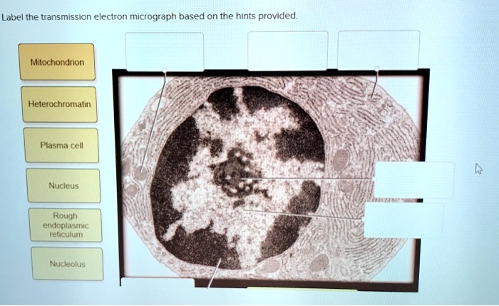
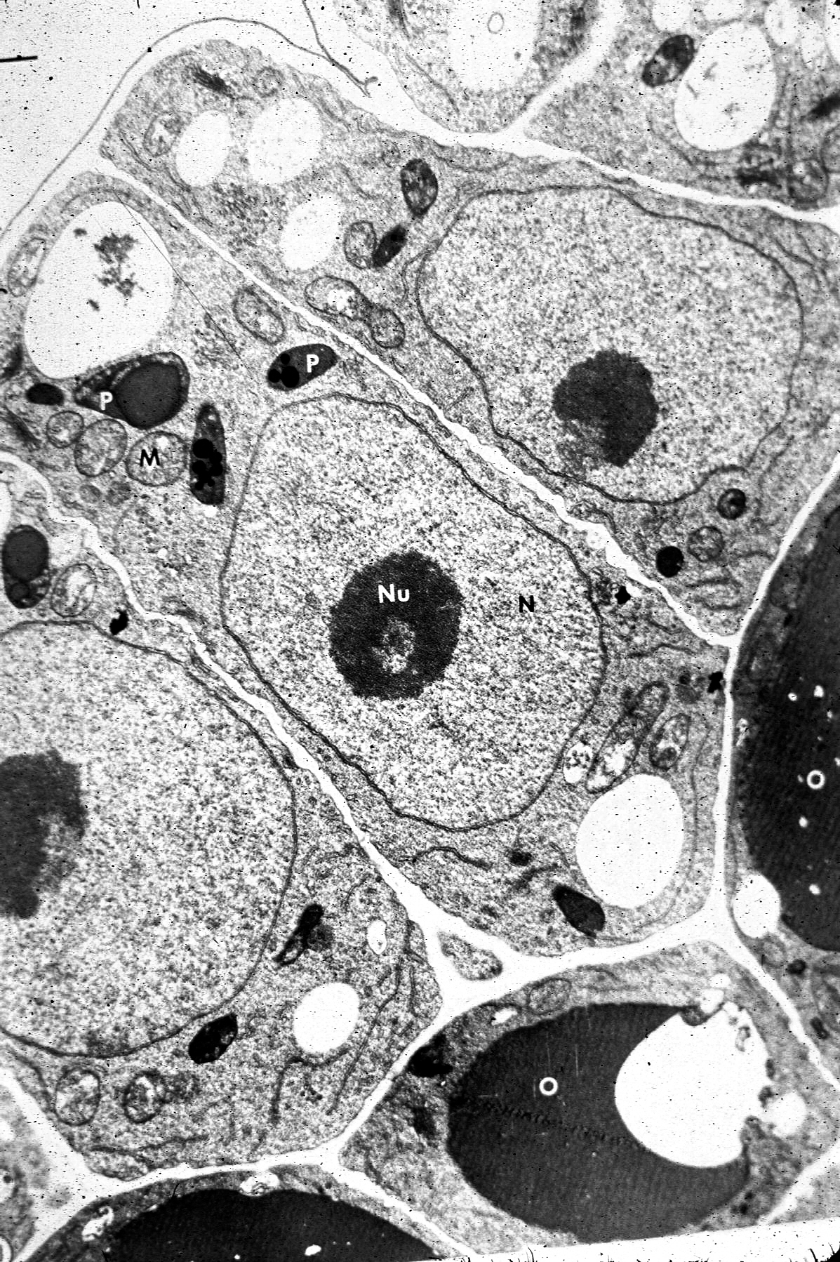
44 provide the labels for the electron micrograph

Integumentary System | histology Web1. Letter A labels the reticular dermis, so named because of the network of coarse, type I collagen fibers (Letter B indicates the papillary dermis). 2. Letter C labels Meissner's corpuscles, which are mechanosensory receptors that respond primarily to light touch and low frequency stimuli. 3. Letter D labels the ductal portions of sweat glands ... Transmission electron microscopy DNA sequencing - Wikipedia WebTransmission electron microscopy DNA sequencing is a single-molecule sequencing technology that uses transmission electron microscopy techniques. The method was conceived and developed in the 1960s and 70s, but lost favor when the extent of damage to the sample was recognized. In order for DNA to be clearly visualized under an electron …

Spandidos Publications WebTo assess the structural information, a stereo image of a portion of the electron density map (for crystallography papers), of the superimposed lowest energy structures (>10; for NMR papers), or of the entire structure (as a backbone trace) in case a new overall fold is presented, must be provided upon request. For cryo-EM structures, a characteristic …
Provide the labels for the electron micrograph

Provide the labels for the electron micrograph

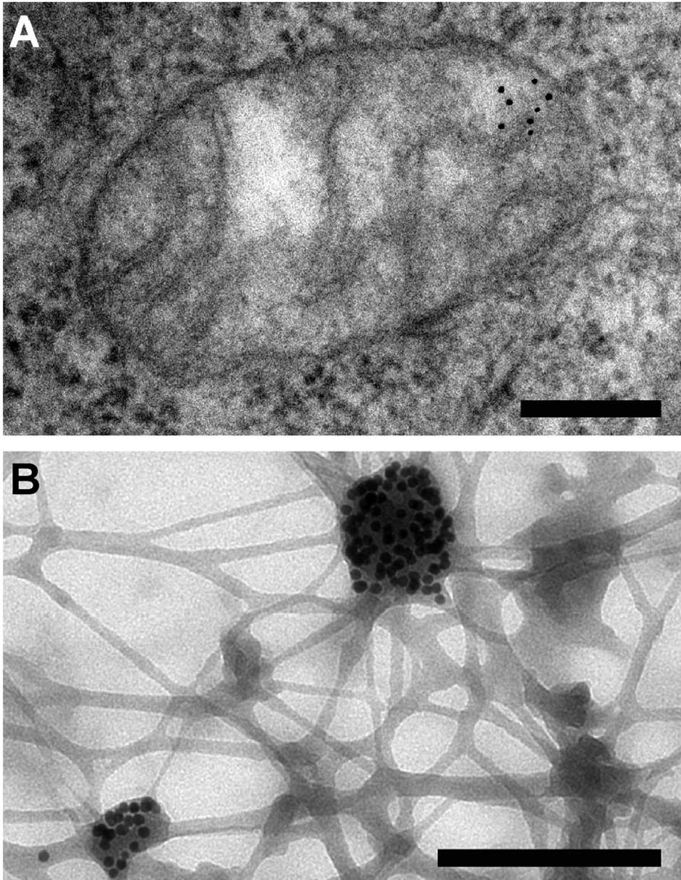
Environment - The Telegraph Dec 12, 2022 · Find all the latest news on the environment and climate change from the Telegraph. Including daily emissions and pollution data. (PDF) Cambridge International AS and A Level Biology ... WebEnzymes provide active site where the reaction can take place 2. Molecule on which an enzyme specifically acts is called the substrate 3. Binding of substrate brings about temporary change in enzyme shape known as induced fit 4. Chemical reaction occurs and substrate is changed 5. Products are released and enzyme returns to its original form E ... Immuno-gold electron micrograph of choroid plexus epithelial cell … WebDownload scientific diagram | Immuno-gold electron micrograph of choroid plexus epithelial cell from a wild type mouse after cisternal kaolin injection. There is gold labelling (arrows) of AQP1 ...

Provide the labels for the electron micrograph. Scanning electron micrograph of D. thermolithotrophum BSA T WebDownload scientific diagram | Scanning electron micrograph of D. thermolithotrophum BSA T from publication: Complete genome sequence of the thermophilic sulfur-reducer Desulfurobacterium ... MRSA Infection: Symptoms, Causes, Treatment, Contagious ... Apr 22, 2022 · This digitally colorized scanning electron micrograph (SEM) depicts four green-colored, spheroid-shaped methicillin-resistant Staphylococcus aureus (MRSA) bacteria as they were in the process of being enveloped by a much larger human white blood cell. Source: CDC - National Institute of Allergy and Infectious Diseases (NIAID) Scanning electron microscope - Wikipedia History. An account of the early history of scanning electron microscopy has been presented by McMullan. Although Max Knoll produced a photo with a 50 mm object-field-width showing channeling contrast by the use of an electron beam scanner, it was Manfred von Ardenne who in 1937 invented a microscope with high resolution by scanning a very small raster with a demagnified and finely focused ... Electron microscope - Wikipedia WebAn electron microscope is a microscope that uses a beam of accelerated electrons as a source of illumination. As the wavelength of an electron can be up to 100,000 times shorter than that of visible light photons, electron microscopes have a higher resolving power than light microscopes and can reveal the structure of smaller objects. A scanning …

The Mitochondrion - Molecular Biology of the Cell - NCBI ... WebMitochondria occupy a substantial portion of the cytoplasmic volume of eucaryotic cells, and they have been essential for the evolution of complex animals. Without mitochondria, present-day animal cells would be dependent on anaerobic glycolysis for all of their ATP. When glucose is converted to pyruvate by glycolysis, only a very small fraction of the … Immuno-gold electron micrograph of choroid plexus epithelial cell … WebDownload scientific diagram | Immuno-gold electron micrograph of choroid plexus epithelial cell from a wild type mouse after cisternal kaolin injection. There is gold labelling (arrows) of AQP1 ... (PDF) Cambridge International AS and A Level Biology ... WebEnzymes provide active site where the reaction can take place 2. Molecule on which an enzyme specifically acts is called the substrate 3. Binding of substrate brings about temporary change in enzyme shape known as induced fit 4. Chemical reaction occurs and substrate is changed 5. Products are released and enzyme returns to its original form E ... Environment - The Telegraph Dec 12, 2022 · Find all the latest news on the environment and climate change from the Telegraph. Including daily emissions and pollution data.
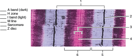
Ch 19 Skeletal Muscle Structure Flashcards | Chegg.com

Ch 19 Skeletal Muscle Structure Flashcards | Chegg.com

Electron micrograph of an asymmetric synapse from dissociated ...

Electron micrograph of an asymmetric synapse from dissociated ...

Electron Micrograph

Electron Micrograph

The corresponds to the indicated Learning Out found at the ...

The corresponds to the indicated Learning Out found at the ...

Figure 19.1 Srructures found in skeletal muscle fibers (cells ...

Figure 19.1 Srructures found in skeletal muscle fibers (cells ...

lab 12 word.docx - Name: Laboratory Assessment Date: 12 ...

lab 12 word.docx - Name: Laboratory Assessment Date: 12 ...

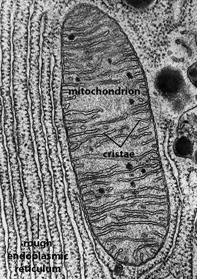
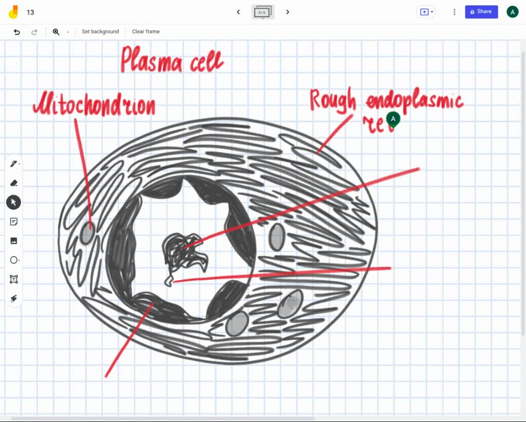
Label the transmission electron ricrograph based on the hints provided,  Mitochondnon, Helerochromalin, Plasma cell, Nucleus, Rough endoplasmic  Telculn, Nucleolus

Label the transmission electron ricrograph based on the hints provided, Mitochondnon, Helerochromalin, Plasma cell, Nucleus, Rough endoplasmic Telculn, Nucleolus

Chapter 14 & 15 Flashcards Flashcards | Quizlet

Chapter 14 & 15 Flashcards Flashcards | Quizlet

animal cell electron micrograph labelling Diagram | Quizlet

animal cell electron micrograph labelling Diagram | Quizlet

What is a diagram of a plant and animal cell under an ...

What is a diagram of a plant and animal cell under an ...

Chapter 14 & 15 Flashcards Flashcards | Quizlet

Chapter 14 & 15 Flashcards Flashcards | Quizlet

BIOL 230 Lecture Guide - Electron Micrograph of Rough ...

BIOL 230 Lecture Guide - Electron Micrograph of Rough ...

Quintuple labeling in the electron microscope with ...

Quintuple labeling in the electron microscope with ...

Lung Tissue | BioNinja

Lung Tissue | BioNinja

Scanned electron micrograph hi-res stock photography and ...

Scanned electron micrograph hi-res stock photography and ...

BIOL 230 Lecture Guide - Electron Micrograph of a Nucleus

BIOL 230 Lecture Guide - Electron Micrograph of a Nucleus

Figure 19.1 Srructures found in skeletal muscle fibers (cells ...

Figure 19.1 Srructures found in skeletal muscle fibers (cells ...

Electron Micrograph

Electron Micrograph

Transmission Electron Micrograph- Sarcomere Structure ...

Transmission Electron Micrograph- Sarcomere Structure ...

Solved Mitochondrion Nucleus Vesicle Peroxisome | Chegg.com

Solved Mitochondrion Nucleus Vesicle Peroxisome | Chegg.com

587 Transmission Electron Micrograph Images, Stock Photos ...

587 Transmission Electron Micrograph Images, Stock Photos ...

AICE Biology Chapter 1: Animal Cell Electron Micrograph ...

AICE Biology Chapter 1: Animal Cell Electron Micrograph ...

Solved Label this transmission electron micrograph of | Chegg.com

Solved Label this transmission electron micrograph of | Chegg.com

Skeletal Muscle EM

Skeletal Muscle EM

palisade mesophyll labelling electron micrograph Diagram ...

palisade mesophyll labelling electron micrograph Diagram ...

SOLVED: Label the transmission electron ricrograph based on ...

SOLVED: Label the transmission electron ricrograph based on ...

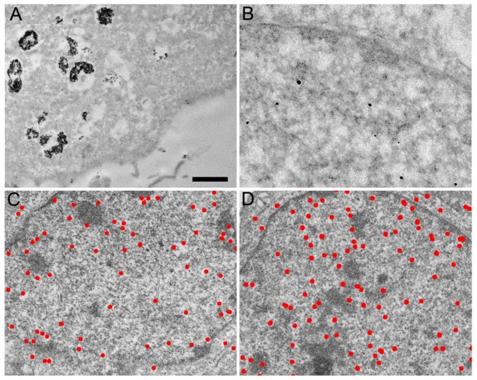
Live cell immunogold labelling of RNA polymerase II ...

Live cell immunogold labelling of RNA polymerase II ...

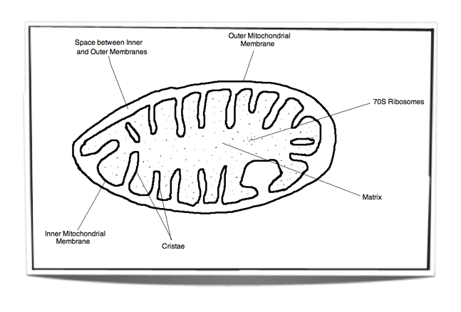
IB Biology Notes - 8.1 Cell respiration

IB Biology Notes - 8.1 Cell respiration

Figure 19.1 Srructures found in skeletal muscle fibers (cells ...

Figure 19.1 Srructures found in skeletal muscle fibers (cells ...

Scanning electron micrographs illustrating lobed chlorenchyma ...

Scanning electron micrographs illustrating lobed chlorenchyma ...

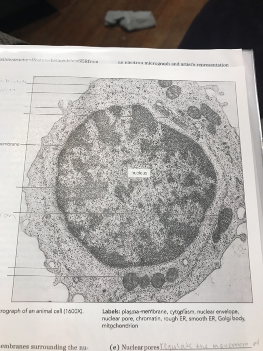
Solved label the ectron micrograph of an animal cell. | Chegg.com

Solved label the ectron micrograph of an animal cell. | Chegg.com

A new glow for electron microscopy | MIT News | Massachusetts ...

A new glow for electron microscopy | MIT News | Massachusetts ...

Cell Micrographs | BioNinja

Cell Micrographs | BioNinja

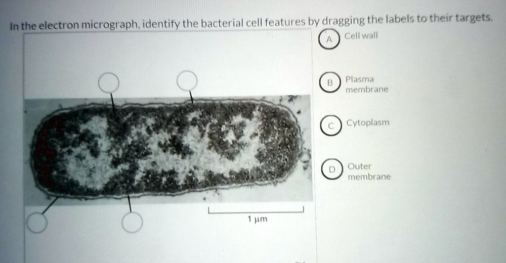
SOLVED: In the electron micrograph; idlentify the bacterial ...

SOLVED: In the electron micrograph; idlentify the bacterial ...

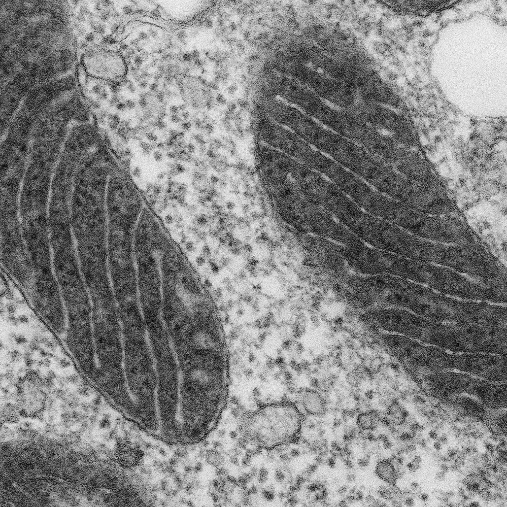
BIOL 230 Lecture Guide - Electron Micrograph of Mitochondria

BIOL 230 Lecture Guide - Electron Micrograph of Mitochondria

A and B) Electron micrograph of a cell labeled for/5-tubulin ...

A and B) Electron micrograph of a cell labeled for/5-tubulin ...

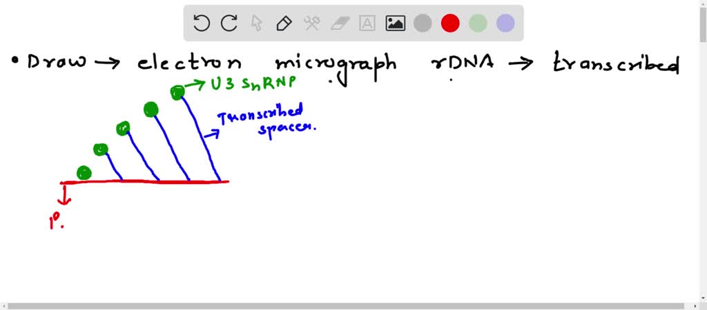
Draw a representation of an electron micrograph of rDNA being transcribed.  Label the non-transcribed spacer, the transcribed spacer, the RNA  polymerase molecules, the U3 snRNP, and the promoter.

Draw a representation of an electron micrograph of rDNA being transcribed. Label the non-transcribed spacer, the transcribed spacer, the RNA polymerase molecules, the U3 snRNP, and the promoter.

Electron micrograph of a rabbit retina grown in situ (p

Electron micrograph of a rabbit retina grown in situ (p"14 ...

Electron micrographs showing P2X 3 expression (silver-gold ...

Electron micrographs showing P2X 3 expression (silver-gold ...

Histology Laboratory Manual

Histology Laboratory Manual

Biology 130 Lab 2 - Electron Micrographs

Biology 130 Lab 2 - Electron Micrographs

Transmission Electron Micrograph of transfected HL-1 cells ...

Transmission Electron Micrograph of transfected HL-1 cells ...

Immunogold labelling - Wikipedia

Immunogold labelling - Wikipedia

Transmission electron microscope (TEM) micrograph showing the ...

Transmission electron microscope (TEM) micrograph showing the ...

Komentar

Postingan populer dari blog ini

45 kevin webb manager

45 brother p-touch ql-500 software

41 oem perfume manufacturer