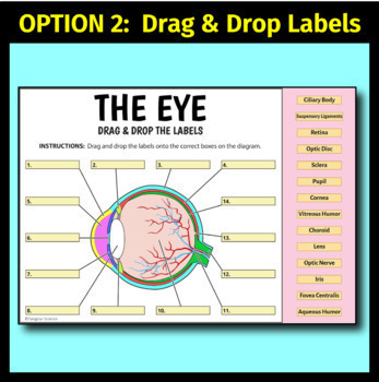
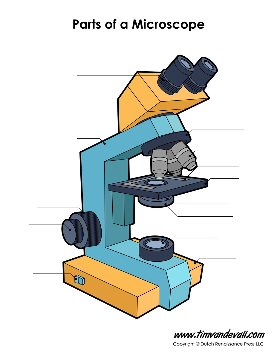
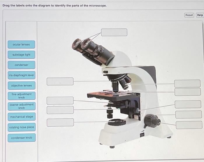
43 drag the labels onto the diagram to identify the parts of the microscope.
compound microscope parts (labeling) Flashcards | Quizlet Created by barnettlily Terms in this set (14) eyepiece tube - connects the eyepiece to the objective lens what is 1? nosepiece (turret) - holds and spins the objective lenses what is 2? 4x objective lens - the 'scanning" objective lens with the lowest magnification what is 3? 10x objective lens - the "low" power objective lens what is 4? Microscope Parts and Functions 5 Hobby Microscopes for Beginners First, the purpose of a microscope is to magnify a small object or to magnify the fine details of a larger object in order to examine minute specimens that cannot be seen by the naked eye. Here are the important compound microscope parts... Eyepiece: The lens the viewer looks through to see the specimen.
Drag The Labels Onto The Diagram To Identify The Structures And ... Drag the labels on the left onto the diagram of the animal cell to correctly identify the function performed by each cellular structure. * fibrous structure around the glenoid fossa. Drag the labels onto the diagram to the stadium wave climate etc. Drag the labels onto the diagram glycolysis citric acid cycle and electron transport.

Drag the labels onto the diagram to identify the parts of the microscope.
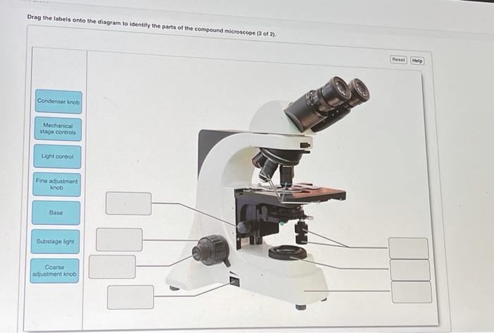
Microsoft takes the gloves off as it battles Sony for its Activision ... 12.10.2022 · Microsoft is not pulling its punches with UK regulators. The software giant claims the UK CMA regulator has been listening too much to Sony’s arguments over its Activision Blizzard acquisition. Drag the labels onto the diagram to identify the part… - SolvedLib Drag the labels onto the diagram to identify the parts and ligaments of the hip joint. Ret Putomom Crostor trochantor Sohead Album A terview with the removed Articular cartage game An anterior to view iconal iments that add strength to the cute Science Biology 0 < Previous Next > Answers Answers #1 Hope it clear your doubt. Thank you Solved Drag the labels onto the diagram to identify the - Chegg Question: Drag the labels onto the diagram to identify the parts of the compound microscope (2 of 2) Malo Condenser knob Mechanical stage controls Light control Fine adjustment knob Base Substage light Coarso adjustmont knob This problem has been solved! You'll get a detailed solution from a subject matter expert that helps you learn core concepts.
Drag the labels onto the diagram to identify the parts of the microscope.. Drag the labels onto the diagram to identify structural features ... Part A Drag the labels onto the diagram to identify structural features associated with a skeletal muscle fiber. ANSWER: Correct HelpReset HelpResetEndomysiumEpimysium Perimysium Nerve Muscle fascicle Blood vesselsMuscle fibers Mitochondria Terminal cisterna Sarcolemma Sarcoplasm Myofibril Thin filaments Triad T tubulesThick filaments ... The Drag The Identify Labels Structures. Diagram To Onto The It carries the optical parts in the upper part of the microscope Feb 22, 2013 · In the video, I demonstrate the movement of a camera in Unity 2021 · drag the labels onto the diagram to identify the structures and ligaments of the shoulder joint Why Is My Left Rib Cage Sore Drag The Labels Onto The Diagram To Identify The Structures Free Body Diagrams, Tutorials with Examples and Explanations ... Click And Drag Each Label To Identify The Organelleslines the … The cell cycle Drag the pink labels onto the pink targets to identify the two main phases of the cell cycle Aug 22, 2018 · Drag the labels onto the diagram to identify the stages in which the lagging strand is synthesized More science games that work on tablets, desktops, laptops & phones organelle that controls all the activities of a cell. Latest Breaking News, Headlines & Updates | National Post Read latest breaking news, updates, and headlines. Get information on latest national and international events & more.
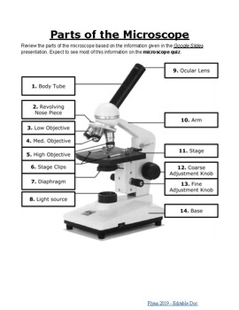
Solved Drag the labels onto the diagram to identify the - Chegg Expert Answer 100% (7 ratings) 1- ocular lens 2-rotating nose piece 3- condenser 4 … View the full answer Transcribed image text: Drag the labels onto the diagram to identify the parts of the microscope. Parts of the Microscope with Labeling (also Free Printouts) A microscope is one of the invaluable tools in the laboratory setting. It is used to observe things that cannot be seen by the naked eye. Table of Contents 1. Eyepiece 2. Body tube/Head 3. Turret/Nose piece 4. Objective lenses 5. Knobs (fine and coarse) 6. Stage and stage clips 7. Aperture 9. Condenser 10. Condenser focus knob 11. Iris diaphragm PlayStation userbase "significantly larger" than Xbox even if … Oct 12, 2022 · Microsoft has responded to a list of concerns regarding its ongoing $68bn attempt to buy Activision Blizzard, as raised by the UK's Competition and Markets Authority (CMA), and come up with an ... Lifestyle | Daily Life | News | The Sydney Morning Herald The latest Lifestyle | Daily Life news, tips, opinion and advice from The Sydney Morning Herald covering life and relationships, beauty, fashion, health & wellbeing
Access Denied - LiveJournal Access Denied - LiveJournal 16 Parts of a Compound Microscope: Diagrams and Video The 16 core parts of a compound microscope are: Head (Body) Arm Base Eyepiece Eyepiece tube Objective lenses Revolving Nosepiece (Turret) Rack stop Coarse adjustment knobs Fine adjustment knobs Stage Stage clips Aperture Illuminator Condenser Diaphragm Video: Parts of a compound Microscope with Diagram Explained Parts of a microscope with functions and labeled diagram - Microbe Notes Q. List down the 18 parts of a Microscope. 1. Ocular Lens (Eye Piece) 2. Diopter Adjustment 3. Head 4. Nose Piece 5. Objective Lens 6. Arm (Carrying Handle) 7. Mechanical Stage 8. Stage Clip 9. Aperture 10. Diaphragm 11. Condenser 12. Coarse Adjustment 13. Fine Adjustment 14. Illuminator (Light Source) 15. Stage Controls 16. Base 17. Science — Biology – Easy Peasy All-in-One Homeschool Lesson 1. Welcome to your first day of school! I wanted to give you one important reminder before you begin. Many of your lessons below have an internet link for you to click on. When you go to the different internet pages for your lessons, please DO NOT click on anything else on that page except what the directions tell you to. DO NOT click on any advertisements or games.
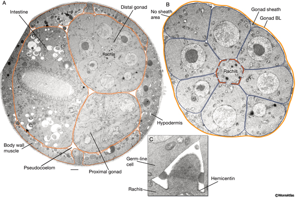
Ultrastructure of cells 1.2
Compound Microscope Parts - Labeled Diagram and their Functions The eyepiece (or ocular lens) is the lens part at the top of a microscope that the viewer looks through. The standard eyepiece has a magnification of 10x. You may exchange with an optional eyepiece ranging from 5x - 30x. [In this figure] The structure inside an eyepiece. The current design of the eyepiece is no longer a single convex lens.

Gravity-resisting colloidal collectives | Science Advances
Labeling the Parts of the Microscope | Microscope activity, Science ... More like this. Bioimager is a professional microscopy company to provide custom microscopy solution for life science and industrial applications at great price & quality. Compound microscope is a microscope that uses multiple lenses to enlarge the image of sample. *This post contains affiliate links. Parts of a syringe (plain tip) and needle.

Mass photometric detection and quantification of nanoscale α ...
Overwatch 2 reaches 25 million players, tripling Overwatch 1 daily ... 14.10.2022 · Following a bumpy launch week that saw frequent server trouble and bloated player queues, Blizzard has announced that over 25 million Overwatch 2 players have logged on in its first 10 days."Sinc

Toward High Spatially Resolved Proteomics Using Expansion ...
Label the microscope — Science Learning Hub Jun 08, 2018 · All microscopes share features in common. In this interactive, you can label the different parts of a microscope. Use this with the Microscope parts activity to help students identify and label the main parts of a microscope and then describe their functions. Drag and drop the text labels onto the microscope diagram. If you want to redo an ...
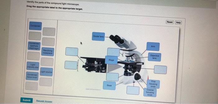
Solved Identify the parts of the compound light microscope ...
The Diagram Onto Identify To Labels Drag Structures. The The So the two pure in bases are adding and guanine Arms - This is the part connecting the base and to the head and the eyepiece tube to the base of the microscope Drag the labels onto the diagram to identify the different intercellular junctions found in animal tissues Skar Audio 10 Inch Subwoofer Box Drag the labels onto the diagram to identify ...

A Survey of Visualization and Analysis in High‐Resolution ...
How to run an assay | Agilent Inspect the cells under the microscope to ensure that cells were not disturbed or washed away. Observe the assay wells under the microscope to ensure that cells were not washed away. Place the plate in a 37° C incubator without CO 2 for one hour prior to the assay. Basic procedures for seeding suspension cells

Chapter 14 - Layers of the Skin - BIO 140 - Human Biology I ...
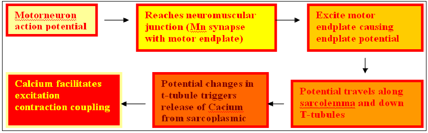
Drag the labels onto the diagram to identify parts of the neuromuscular ... Jan 3, 2022 - Drag the labels onto the diagram to identify parts of the neuromuscular junction. - Label the parts of the neuromuscular junction. Drag the labels onto the

Drag the labels onto the diagram to identify the gustatory ...
Parts of a Microscope - The Comprehensive Guide Step 1: Fully open field and condenser diaphragms and focus on specimen using x10 objective. Step 2: Fully close field diaphragm and adjust the condenser and focus so edges are as sharp as possible. Step 3: Use screws at front of condenser to centre field diaphragm and open field diaphragm to fill view. Step 4: Remove eyepiece and close down ...
ashishware.com
a&p lab 3 hw Flashcards | Quizlet Drag the labels onto the diagram to identify the parts of the compound microscope (1 of 2). (figure 3.1) left column: arm mechanical stage right column: ocular lense ... Recall from the video the parts of a typical compound microscope. Drag the labels to identify the parts of the compound microscope. Not all labels will be used. 1. left column ...

Protocol for intervention-free quantification of protein ...
drag the labels onto the diagram to identify the structures Web Drag the labels onto the diagram to identify the structures and ligaments of the shoulder joint. Web Part a drag the labels onto the diagram to identify the melanocyte in the. Web Thick skin lacks. First drag pink labels to pink targets to label the. Musculotendinous cuff the circle of tendons around the shoulder joint.
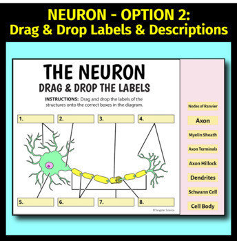
Neuron & Synapse - Google Slide Activities | Distance Learning | 10 Options
Bio2514 Week 3 The Microscope - Lab Topic.docx - Course Hero Drag the labels onto the diagram to identify the parts of the compound microscope (1 of 2). Arm ocular lens Mechanical stage rotating nose piece Stage Objective lenses Condenser Iris diaphragm lever 3. The microscope slide rests on the __________ while being viewed. Stage 4. Your lab microscope is parfocal.
Anatomy and Physiology Lab I” on OpenALG
Identify The Diagram Drag The To Structures. Labels Onto The Drag the labels onto the diagram to identify the types of bone cells Unity drag object with mouse Drag the with resolution 2232px x 1472px Drag the labels onto the diagram to identify the bones and parts of the hip bone In addition to reviewing the human heart anatomy, we will also discuss the function and order in which blood flows through the ...
Microscope Labeling Teaching Resources | Teachers Pay Teachers
PPIC Statewide Survey: Californians and Their Government Oct 26, 2022 · Key Findings. California voters have now received their mail ballots, and the November 8 general election has entered its final stage. Amid rising prices and economic uncertainty—as well as deep partisan divisions over social and political issues—Californians are processing a great deal of information to help them choose state constitutional officers and state legislators and to make ...
Eye & Retina - Google Slide Activities | Distance Learning | 16 Options
Drag The Labels Onto The Diagram To Identify The Structures And ... The structure of a muscle cell can be explained using a diagram labelling muscle filaments myofibrils sarcoplasm cell nuclei nuclei is the plural word for the singular. Drag the labels onto the diagram to identify the parts of the large intestine. The transverse humeral ligament is not shown on this diagram.
Microscopy
10+ drag the labels onto the diagram to identify the structures Drag The Labels Onto The Diagram To Identify The Structures And Ligaments Of The Shoulder Joint. This diagram shows the bones of the upper arm and the elbow joint. Excess plasma lipids in the form of cholesterol contribute to the formation of atherosclerotic plaques.
Microscope Diagram Labeled, Unlabeled and Blank | Parts of a ...
Drag the labels onto the diagram to identify parts of the neuromuscular ... Drag the labels onto the diagram to identify parts of the neuromuscular junction. Label the parts of the neuromuscular junction. Drag the labels onto the diagram to identify parts of the neuromuscular junction. Label the parts of the neuromuscular junction.

Molecular biology software - CB-MAS - CapitalBio Technology ...
Drag The Labels Onto The Diagram To Identify The Structures And ... Drag the labels onto the diagram to identify the parts of the renal corpuscle. You can click to make it bigger! 19 2 Bone Concepts Of Biology 1st Canadian Edition from opentextbc.ca An example of a synchondrosis is the articulation of the.
Quizbank/All questions - Wikiversity
Solved Drag the labels onto the diagram to identify the - Chegg Question: Drag the labels onto the diagram to identify the parts of the compound microscope (2 of 2) Malo Condenser knob Mechanical stage controls Light control Fine adjustment knob Base Substage light Coarso adjustmont knob This problem has been solved! You'll get a detailed solution from a subject matter expert that helps you learn core concepts.
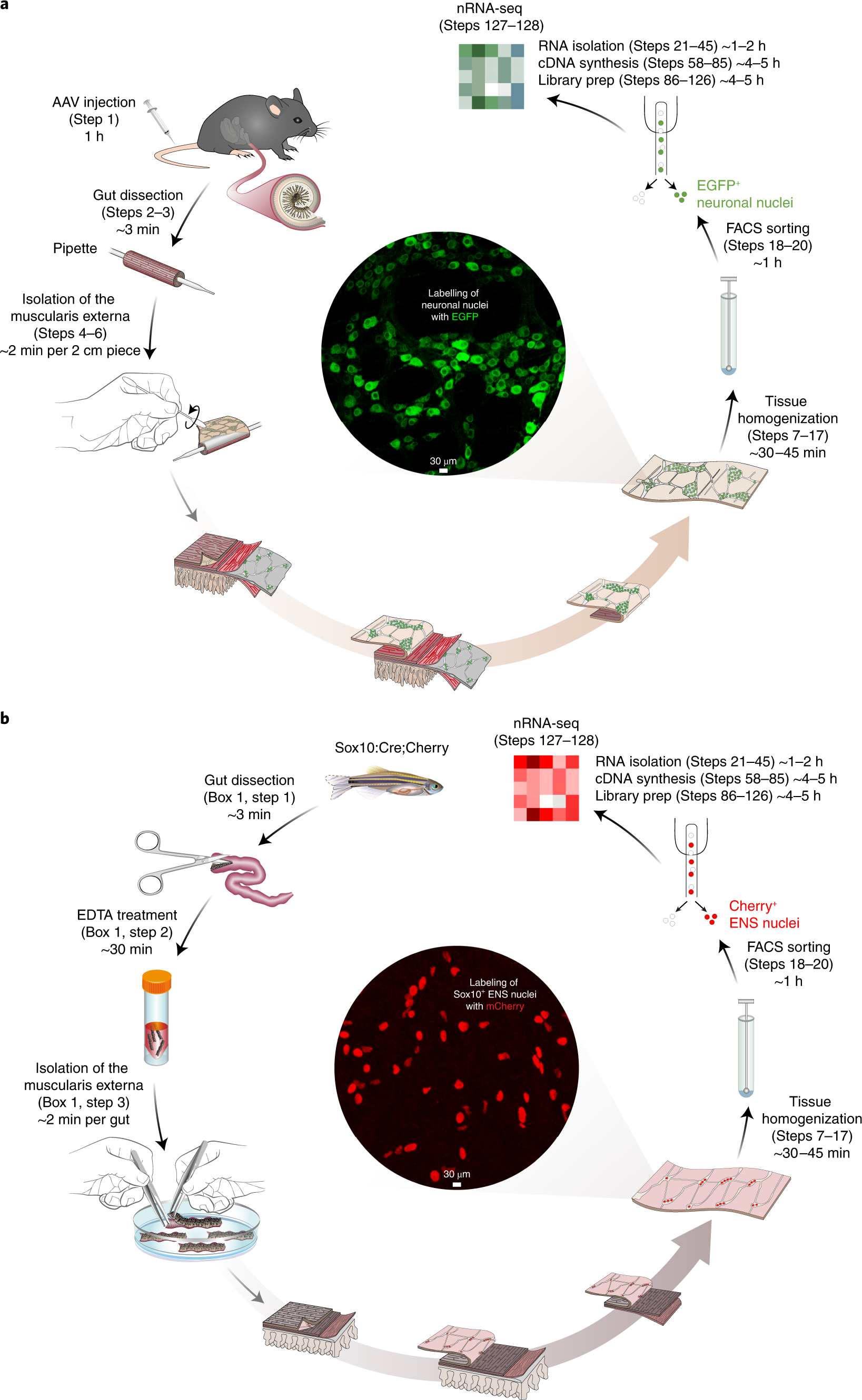
Molecular profiling of enteric nervous system cell lineages ...
Drag the labels onto the diagram to identify the part… - SolvedLib Drag the labels onto the diagram to identify the parts and ligaments of the hip joint. Ret Putomom Crostor trochantor Sohead Album A terview with the removed Articular cartage game An anterior to view iconal iments that add strength to the cute Science Biology 0 < Previous Next > Answers Answers #1 Hope it clear your doubt. Thank you
What are some of the most interesting biological organelles ...
Microsoft takes the gloves off as it battles Sony for its Activision ... 12.10.2022 · Microsoft is not pulling its punches with UK regulators. The software giant claims the UK CMA regulator has been listening too much to Sony’s arguments over its Activision Blizzard acquisition.
QuickFigures: A toolkit and ImageJ PlugIn to quickly ...

Microscopy
ZEISS LSM 880 OPERATING MANUAL Pdf Download | ManualsLib

Between the (Gender) Lines: the Science of Transgender ...

Label the microscope — Science Learning Hub

Chemical Analysis of Microplastics and Nanoplastics ...
Answered: Drag the appropriate labels to their… | bartleby
Solved Drag the labels onto the diagram to identify the ...
Solved Drag the labels onto the diagram to identify the ...

Simple and selective paper-based colorimetric sensor for ...

The Scientist Who Scrambled Darwin's Tree of Life - The New ...

Microbiology Final Flashcards | Quizlet

Characterization of nanoparticles for therapeutics | Nanomedicine
Handbook - Pericellular Structures

Semiotics for Beginners by Daniel Chandler
Unit 3

Phage Hunt NZ | Massey University Phage Hunters
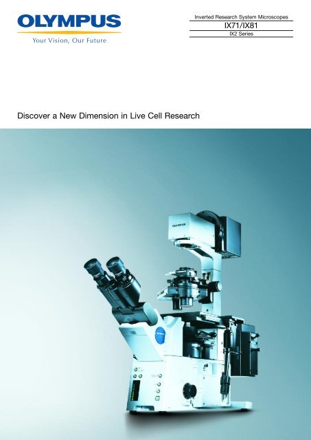
Olympus IX81 Brochure - Microscopes

Answered: a the labels onto the diagram to… | bartleby
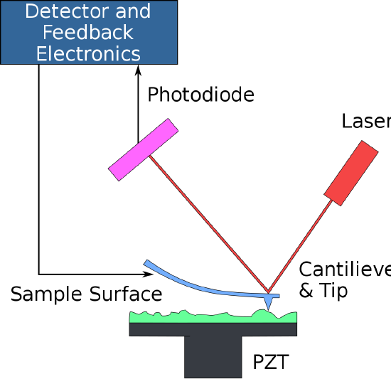
9.2: Atomic Force Microscopy (AFM) - Chemistry LibreTexts

Structure and Function of Blood Vessels - Human Biology ...

Chapter: The Cell — The Biology Primer
Costus stripe mosaic virus, a tentative new member of the ...


Komentar
Posting Komentar